Intro
Gallery
Video


General Overview
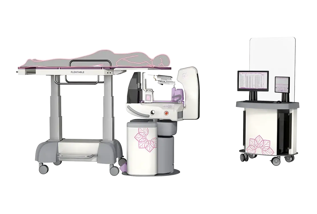
Advanced and innovative breast tomosynthesis system including the integrated capability to generate full-field digital mammography (FFDM) images.
It features a unique and ergonomic design which ensures patient comfort and easiness for the operator: the gantry is able to reach different positions, including the horizontal one for interventional procedures. A high-precision tomo-guided biopsy kit allows the operator to reduce procedure time and patient anxiety.
The system is equipped with dedicated algorithms for optimal image quality, user-friendly Raffaello software’s interface and safe diagnosis even with Dual Energy technique (CESM).
Ceramic X-ray tube

High performance generator
Tilting movements
Prone biopsy

Raffaello software

Tomosynthesis (DBT)

Dual Energy (CESM)

Dose Reduction

Download Brochure
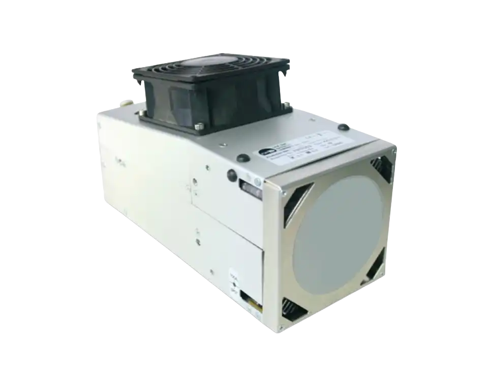
Ceramic X-ray tube
The special metal-ceramic X-ray tube guarantees continuous heat dissipation for high patient’s throughput and high performance in all acquisition modalities, while assuring lower maintenance practices.
It features a reduced weight and a higher stability to perform a Step&Shoot DBT acquisition in order to get images with more sharpness and details.

High performance generator
Viola DBT is characterized by high-performance generators able to reach powers up to 8 kW, reducing exposure time during CESM exams of thicker breast tissues.
Tilting movements
The innovative design of the gantry enables a wide freedom of quick movements in any direction.
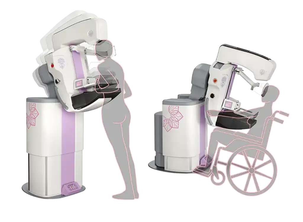
The forward tilting movement (up to -10°) allows the positioning of non-collaborative patients (i.e., in wheelchair), while the backward tilting movement (up to +15°) ensures the relaxation of the pectoral muscle in order to include more breast tissue into the view.
Furthermore, Viola DBT is the only system able to reach the horizontal position (+90°), performing prone biopsies with the dedicated Flexitable.
Prone biopsy
Thanks to the unique rotating movement up to horizontal position and the dedicated Flexitable, radiologists can perform prone breast biopsies with higher confidence and better patient comfort.
- Smart Finder, used for stereotactic, tomo-guided or combined biopsies, is a high-precision needle simulator software which drives to choose the best approach to the lesion;
- Flexitable is manoeuvrable and ergonomic, with reduced weight, special wheels with electromechanical brakes, adjustable breast aperture and possible inclination;
- Smart Checker is an optional biopsy accessory device which arranges samples of biopsy, allowing the acquisition of images of the tissue cores on the uncovered part of the detector’s active area, without any mammary decompression.
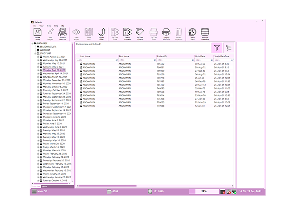
Raffaello software
Raffaello, the dedicated management software for breast care, ensures a rapid and easy workflow for each acquisition modality (2D, DBT, BIOPSY and CESM).
The user-interface is easy and intuitive and it is configurable according to the operator’s needs.
The special Acquisition Workstation (AWS) allows the visualization during biopsy procedures.
Raffaello software can also be installed on the optional Review Workstation (RWS) with a set of dedicated tools for rapid mammography and tomosynthesis image reviewing.
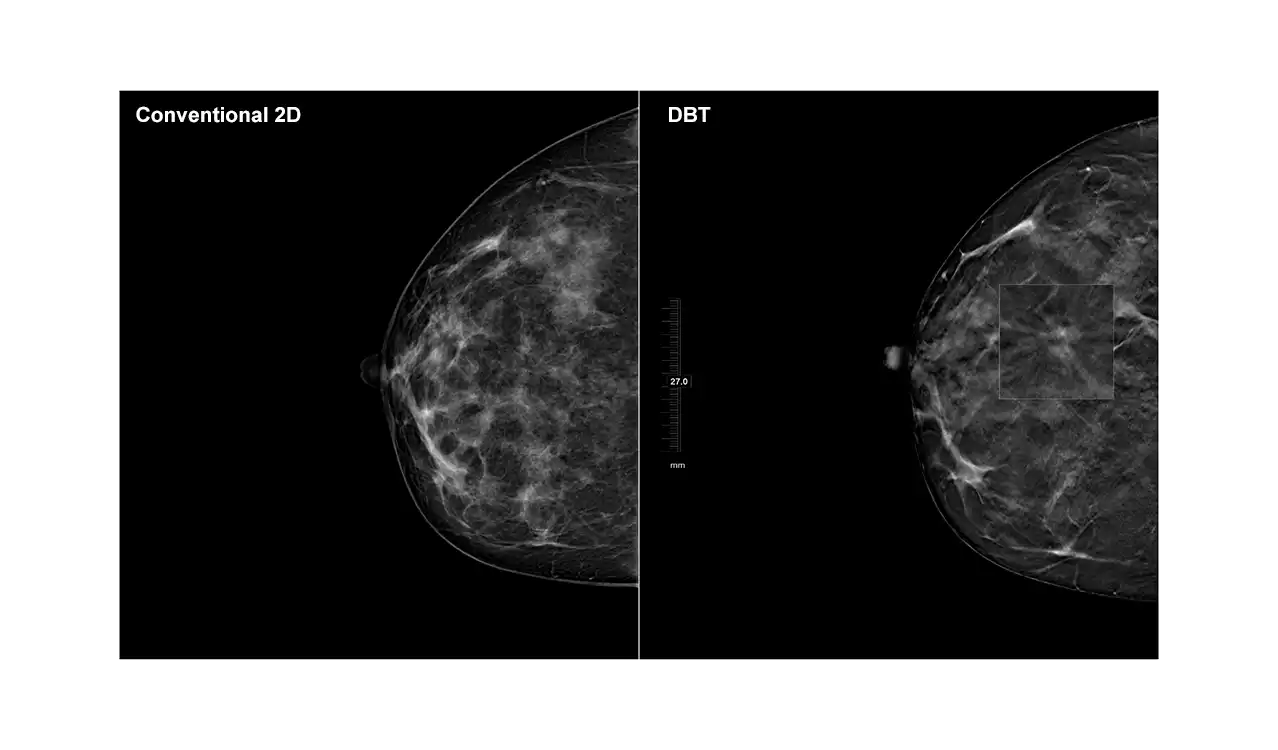
Tomosynthesis (DBT)
Digital Breast Tomosynthesis (DBT) gives an improvement in detection and characterization of lesions.
Step&Shoot acquisition allows exposures while the tube is stationary: the result is an image that is completely devoid of blurring effects and with sharper outlines.
Thanks to the Iterative Reconstruction Algorithm, an extremely fast and accurate tomosynthesis reconstruction with no binning, images have high level of detail, allowing the identification of microcalcifications and tiny breast structures.
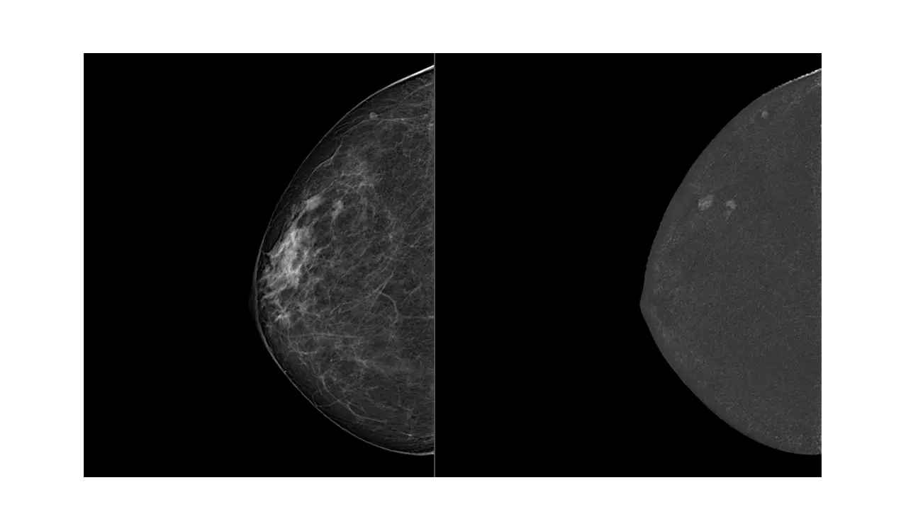
Dual Energy (CESM)
Contrast Enhanced Spectral Mammography (CESM) is a new breast imaging technique. Following the injection of a contrast agent, in a very short time and within a single breast compression, two images are acquired, one with low-energy and one with high-energy. An innovative image subtraction algorithm generates a clinical image which reveals the potential tumour angiogenesis alone.

Dose Reduction
Technologically advanced components and cutting-edge image processing software ensure high image quality with the lowest patient dose in all operating conditions.